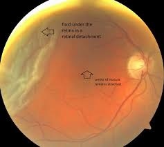
[vc_row][vc_column][vc_single_image source=”external_link” external_img_size=”800×800″ alignment=”center” custom_src=”https://drasifkhokhar.com/Images/Retina/Retina.jpg” title=”Retina is like a lamination on the inner surface of the eye. Small nerve fibers arise from throughout thr retina nad converge in the center at the point which is called optic disc. When we examine with different instruments, retina looks like the pictures below”][vc_single_image source=”external_link” external_img_size=”800×800″ alignment=”center” custom_src=”https://drasifkhokhar.com/Images/Retina/RD1.jpg” title=”The innermost layer seen in the picture is retina”][vc_single_image source=”external_link” external_img_size=”800×800″ alignment=”center” custom_src=”https://drasifkhokhar.com/Images/Retina/RD2.jpg” title=”Holes develop in the retina or sometimes some part of retina torns out. The holes and breaks appear as seen in following picture”][vc_single_image source=”external_link” external_img_size=”800×800″ alignment=”center” custom_src=”https://drasifkhokhar.com/Images/Retina/RD3.jpg” title=”When a part of retina tears out or a hole develops in some part, then fluid perculates and accumulates under the retina. That part of the retina bulges inwards as seen in the pictures”][vc_single_image source=”external_link” external_img_size=”800×800″ alignment=”center” custom_src=”https://drasifkhokhar.com/Images/Retina/RD6.jpg” title=”Detached retina and fluid appears as seen in this picture”][vc_single_image source=”external_link” external_img_size=”800×800″ alignment=”center” custom_src=”https://drasifkhokhar.com/Images/Retina/PPV3.jpg” title=”When retina is detached, one method of reataching it is buckling. A silicon band is tied around the eye which presses from outside and compells the two layers of retina remain attached. The silicon band is seen as following after operation”][vc_single_image source=”external_link” external_img_size=”800×800″ alignment=”center” custom_src=”https://drasifkhokhar.com/Images/Retina/PP4.jpg”][vc_column_text]
The latest method for treatment of retinal detachment is vitrectomy operation. The fluid is drained out and the hole or break is welded by layer. This method is really a miracle. We should be thankfull to the reasearchers and Allah for this blessing to humanity. In the past This operation used to be very lenthy. Sometimes even 14 hours took one operation! bu now due to newer invenstions sometimes can be completed even less than a hour!
[/vc_column_text][vc_single_image source=”external_link” external_img_size=”800×800″ alignment=”center” custom_src=”https://drasifkhokhar.com/Images/Retina/PP5.jpg” title=”You can see how instruments work during vitrectomy operation”][vc_single_image source=”external_link” external_img_size=”800×800″ alignment=”center” custom_src=”https://drasifkhokhar.com/Images/Retina/PPV6.jpg”][vc_single_image source=”external_link” external_img_size=”800×800″ alignment=”center” custom_src=”https://drasifkhokhar.com/Images/Retina/PPV7.jpg”][vc_single_image source=”external_link” external_img_size=”800×800″ alignment=”center” custom_src=”https://drasifkhokhar.com/Images/Retina/PPV8.jpg” title=”Laser is being applied during vitrectomy operation”][vc_single_image source=”external_link” external_img_size=”800×800″ alignment=”center” custom_src=”https://drasifkhokhar.com/Images/Retina/PPV.jpg” title=”I am performing vitrectomy operation by The Storz Company’s Millinuim Vectrectomy Machine, Zeiss Company’s Lumera Model Operation Microscope, and BIOM. This modern method requires neither general anesthesia nor stitching”][vc_single_image source=”external_link” external_img_size=”800×800″ alignment=”center” custom_src=”https://drasifkhokhar.com/Images/Retina/PPV9.jpg” title=”I am performing vitrectomy operation by Oertli Company’s Feros microsurgical system machine, Zeiss Company’s Lumera Model Operation Microscope, BIOM. This modern method requires neither general anesthesia nor stitching.”][/vc_column][/vc_row]
Leave a Reply